

Radiology
Radiology
Radiology, also known as diagnostic imaging, is a series of tests that take pictures or images of parts of the body. The field encompasses two areas — diagnostic radiology and interventional radiology — that both use radiant energy to diagnose and treat diseases. While there are several different imaging exams, some of the most common include x-ray, MRI, ultrasound, CT scan and PET scan.
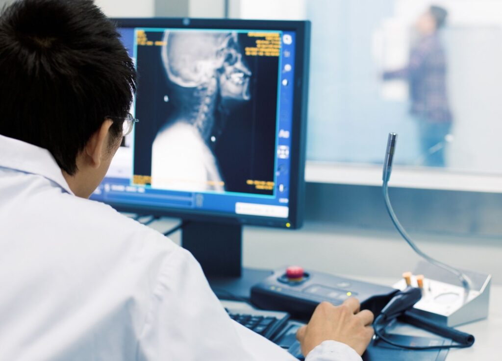
A radiologist will look at the outcome of a certain imaging test to find a relevant image that evaluates and supports a diagnosis. These individuals are usually medical doctors (MDs) with highly specialized training focused on the interpretation of medical imaging. Radiologic technologists also aid in this process, as they use and manage the machines in the course of producing an image. After a patient undergoes imaging tests, radiologists will give reports of their interpretations to the referring clinical doctors.
Radiology Services in Nashik is a branch of medicine concerned with the use of radiant radiation in the diagnosis and treatment of diseases. This discipline is divided into two major categories: diagnostic radiology and interventional radiology. A radiologist is a physician who focuses in radiology.
The outcome of an imaging investigation is not solely dependent on the indication or the technical execution. Diagnostic radiology specialists are the concluding link in the diagnostic chain, searching for pertinent image information to evaluate and ultimately support a sound diagnosis.
Diagnostic Radiology Procedures
There are several ways for obtaining images to aid in the screening, diagnosis, or monitoring of medical conditions. These are some examples:
X-Rays
- X-rays or plain radiographs are frequently used to examine bones, the thorax, or the abdomen. Denser structures, such as bones, appear white (opaque) on X-rays, whereas air-filled regions, such as the lungs, appear black. Most body components are a shade of grey between these two.
- X-rays can be used alone to identify fractures, some pneumonias, and bowel obstructions. However, additional imaging tests are frequently required.
- For example, while chest X-rays can sometimes detect lung cancer, one study2 found that 20% to 23% of these tumors were missed in individuals with lung cancer symptoms (And thus, CT scans are needed for lung cancer screening). Some fractures (such as stress fractures) may require an MRI to be detected.
- The region of the body under investigation may limit the efficacy of X-rays. An abnormality is less likely to be evident on an X-ray of the forearm in areas where several structures overlap (for example, the collar bone, heart, and lung on the left side of the thorax).
- Screening for specific conditions may require the use of specialized X-ray methods. Digital mammography, for example, is an X-ray method that uses low dose radiation to detect breast cancer, whereas panoramic X-rays are used to detect dental disease.
CT (Computerized Tomography) (CT)
- Computed axial tomography (CAT scans or CT scans) creates a cross-sectional image of the inside of the body using a sequence of X-rays and a computer. CT imaging is more detailed than X-ray imaging and can better identify areas where tissues overlap. CT scans can identify smaller abnormalities than traditional X-rays can.
- Contrast dyes used in CT scans can enhance visualization in some areas, such as the digestive tract. CT procedures, such as CT angiography, can provide information that would otherwise require a more invasive operation in some cases.
Magnetic resonance imaging (MRI)
- Magnetic resonance imaging (MRI) is a form of imaging that employs a magnetic field. (MRI)
- Magnetic resonance imaging creates images of the inside of the body by combining powerful magnetic fields and radio waves. While CT is frequently a better method for evaluating bones and blood vessels, MRI is frequently a better method for assessing soft tissue such as the brain, spinal cord, nerves, muscles, tendons, and breast tissue.
- With brain, spinal cord, and peripheral nerve disorders, MRI has enabled healthcare practitioners to diagnose conditions that were previously only clinically assumed. For example, practitioners can now use an MRI to diagnose multiple sclerosis, a diagnosis that was previously confined to an assessment of symptoms alone. MRI is more accurate than mammography for breast cancer screening, but the higher cost makes it impractical for individuals who do not have underlying risk factors for breast cancer (a significant family history of cancer, a BRCA mutation, or a history of childhood cancer). A newer technique known as fast MRI is a quick and low-cost test that may be more effective in detecting early breast cancer in the future.
- Most imaging methods, with the exception of PET/CT (see below), are structural but not functional. This means that they reveal the structure of a body part but do not provide knowledge about its function. However, one type of MRI, known as functional MRI, can provide an indication of brain activity.
- Contrast, like CT, is frequently used to better define regions being scanned, with gadolinium being a popular agent. At times, magnetic resonance technology may be used as an alternative to more invasive treatments, such as magnetic resonance angiography (MRA).
- MRI has the benefit of not using ionising radiation, which has been linked to an increased risk of cancer, particularly in children. The cost, body mass index (MRI is challenging in very overweight people), and the fact that it may not be used in people who have metal in their bodies are three limitations.
Ultrasound
- Ultrasound employs sound waves (acoustic energy) to create moving pictures of a body part. Ultrasound is best recognised as a method for examining a foetus during pregnancy, but it can also assist with some medical conditions.
- Breast ultrasound can frequently differentiate cysts from masses in the breast. Cysts can be aspirated using ultrasound guidance, and their disappearance can also be comforting (no further evaluation may be needed). Heart ultrasound (echocardiogram) can be used to assess heart valves, cardiac motion, the pericardium (heart lining), and more. This technique can be performed by either placing a transducer on the skin overlying the heart or by threading a transducer into the oesophagus (transesophageal echocardiogram).
- Thyroid ultrasonography can be used to determine the presence of thyroid nodules. Abdominal ultrasound is frequently used to detect gallstones and other medical problems.
- Pelvic ultrasonography is frequently used to detect ovarian cysts. Because ultrasound does not use radioactivity, it is safe to use during pregnancy. It is less useful in distinguishing situations where there is no difference in tissue density because it is reliant on finding contrast (such as between a solid mass and a fluid-filled mass).
Fluoroscopy
- Fluoroscopy creates moving pictures of the body by using X-rays in real time. These real-time images are especially essential in some situations.
- Fluoroscopy, for example, may be used to observe changes in contrast flow in joints associated with various movements, in the digestive system with an upper gastrointestinal or barium enema study, or to monitor progress during pacemaker insertion.
- Fluoroscopy's radiation exposure is considerably greater than that of conventional X-rays due to continuous monitoring (multiple images taken over time).
Radiation Medicine Examinations
Nuclear medicine imaging employs radioactive material ("radioactive tracers") that is detected by a camera to create images of the inside of the body. While most imaging techniques are structural in nature, describing structures on the inside of the body, these scans are used to assess how different parts of the body work. The radioactive substance may also be used to treat cancer in some instances (such as the use of radioactive iodine to treat thyroid cancer).
Imaging Molecular
The use of additional specialist methods known as molecular imaging is also possible. This covers processes like dual-energy CT, laser imaging, and CT perfusion.
Interventional Radiology Procedures
Interventional radiology treatments now come in a wide variety. These “minimally intrusive” techniques frequently take the place of more invasive treatments that were previously used, like surgery.
Because of this, these procedures might be less risky, require smaller cuts, be more comfortable for patients, and hasten their recovery times. They frequently cost less. The following is a summary of some of the ailments that could be handled in this manner.
- Detecting and Opening a Restricted Blood Vessel
- Interventional procedures may be used to address blood vessels (either arteries or veins) that are blocked in the heart, legs, or lungs.
- Coronary artery blockages: Coronary artery narrowing or blockages can be addressed with angiography, angioplasty, and stent placement. A wire is introduced into the artery, and a balloon is used to open the narrowing in the artery. In order to open the artery, a clot-busting drug may be injected instead.
- A stent may then be inserted to keep the artery open and enable blood to flow to a damaged area of the heart. If an artery in the heart (heart attack) or limbs becomes suddenly blocked, clot-blasting medication may be injected to open the artery, followed by stent placement if necessary.
- Deep venous thrombosis (blood clots in the veins of the legs or pelvis): If clot-blasting medication (thrombolytics) is discovered, it may be injected through a catheter placed in a vein with the aid of imaging. The insertion of a balloon or stent may then be used.
- Stents can also be used to treat blood vessels that have been compressed by a tumour and are causing problems.
- Pulmonary emboli develop when blood clots (deep vein thromboses) break off in the legs or pelvis and travel to the lungs (pulmonary emboli). A radiologist may occasionally introduce a catheter into an artery to break up a large clot in the lungs.
- A radiologist may also place a filter into the large blood vessel returning blood to the heart for individuals who have recurrent clots in their legs (the inferior vena cava). In this situation, the filter may help to avoid pulmonary emboli.
- Blocking a Blood Artery
- Interventional radiology may also be used to obstruct a vessel. Varicose veins can be treated with vein ablation, whereas fibroids can be treated with artery embolization (uterine artery embolization).
- Aneurysm Therapy : Aneurysms are dilated and weak portions of an artery that are prone to rupture or bleeding. A radiologist may use interventional radiology to put a stent graft in the area of an aneurysm, effectively relining the blood vessel.
- To Stop the Bleeding : Interventional radiology may be used as an alternative to surgery to manage bleeding (hemorrhage) in conditions varying from gastrointestinal bleeding to postpartum bleeding to trauma. Bleeding can be managed by blocking a blood vessel (as mentioned above), inserting a stent, applying pressure with a balloon, and other methods.
- Central Line Positioning : Rapid access to larger blood vessels for infusion is required when a person is critically ill or will be getting caustic medications such as chemotherapy. (Peripheral veins, such as a vein in the hand or forearm, are often insufficient.) Ports and PICC lines are examples of center lines.
- Feeding Tube Positioning : Feeding tube placement (gastrostomy, jejunostomy) is a relatively frequent interventional radiology procedure. When a person is unable to consume for any reason, these are frequently used.
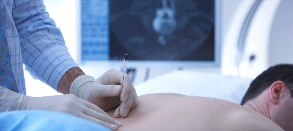
- Tissue Biopsies A radiologist can conduct a variety of biopsy procedures, which are frequently guided by ultrasound or CT. Needle biopsies and guided biopsies are two examples.
- Cancer Therapy :Aside from radiation treatment (discussed further below), a variety of interventional radiology techniques may be used to treat a primary tumor or metastases. (cancer that has spread).
- Tumors can be treated with ablative therapies (tumor-destroying procedures) such as radio frequency ablation or microwave ablation, or by tumor embolization. (blocking a blood vessel that feeds a tumor so that the tumor dies).
- Alternatively, chemotherapy or radiation can be delivered straight to a tumor or metastasis site (chemoembolization/radioembolization).
- For Vertebral Fractures :To address collapsed vertebrae, procedures known as vertebroplasty or kyphoplasty can be used. The radiologist injects a cement-like substance into the fracture to successfully repair it.
- To Resolve Blockages : An interventional radiologist may use a stent to treat obstructions in various parts of the body. This may be done to clear a clogged esophagus, clogged bile ducts, a clogged ureter draining from the kidney, or a clogged intestine.
- Drainage : When fluid accumulates in a body area, an interventional radiologist may place a drain to remove the fluid or pus. This could be done to drain recurring pleural effusions (fluid accumulation around the lungs), in the brain (shunting), and much more.
- Back Pain Treatment Methods : Radiologists now use a variety of procedures to address chronic back pain.
FAQ
Radiology is a medical specialty that uses imaging techniques, such as X-rays, CT scans, MRI, and ultrasound, to diagnose and treat diseases and injuries. Radiologists are medical doctors who specialize in interpreting medical images and providing diagnostic reports to referring physicians.
Radiology employs a number of imaging techniques to visualise various parts of the body, including X-rays, CT scans, MRI, ultrasound, and nuclear medicine. X-rays use electromagnetic radiation to create pictures of the body's internal structures, whereas CT scans use X-rays to create detailed, cross-sectional images of the body. A magnetic field and radio waves are used in MRI to generate images of soft tissues and organs. Ultrasound creates pictures of the body's internal structures by using high-frequency sound waves. Nuclear medicine employs radioactive compounds to visualise organ function and metabolic activity.
The procedures performed in radiology will depend on the type of imaging technique being used. X-rays and CT scans involve lying on a table while the images are taken. MRI involves lying on a table that moves into a large, cylindrical magnet. Ultrasound involves applying a gel to the skin and moving a small device called a transducer over the area being imaged. Nuclear medicine involves the injection of a radioactive substance, followed by imaging of the area of interest. During all imaging procedures, the radiology technologist will provide instructions on how to position your body to obtain the best images. After the procedure, the images will be reviewed by a radiologist, who will provide a report to your referring physician.